Introduction
Histology Core at BRI
The Histology Core Laboratory provides comprehensive histological services to both BRI scientists and the surrounding biotech community, from external academic investigators to large multinational biotech companies.
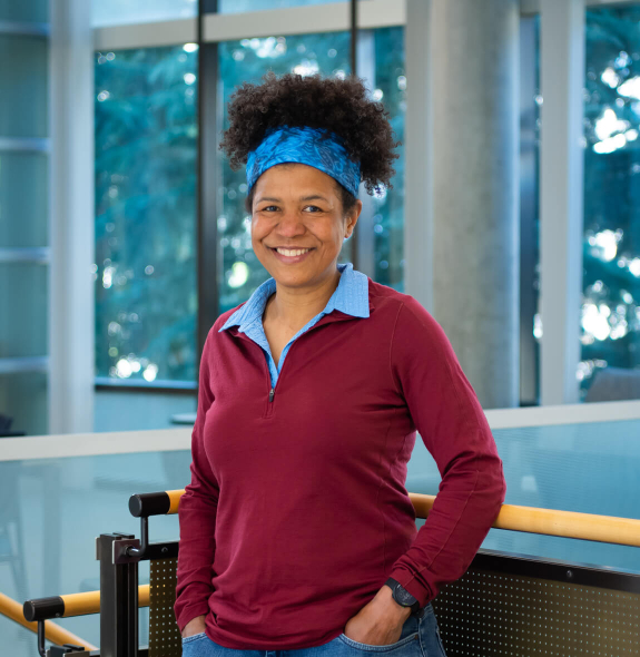
Managed by Dr. Pamela Johnson, the core facility utilizes high-throughput automated equipment for tissue infiltration, embedment in paraffin or OCT, sectioning (microtomy, cryotomy), dye-based and immunohistochemical staining, spatial transcriptomics, tissue matrix array, and cover-slipping. Leveraging Dr. Johnson’s 25+ years’ experience, the facility also specializes in troubleshooting and protocol development.

Histology Core Equipment

Biocare Medical INTELLIPATH FLX®
An automated high throughput immunohistochemistry stainer that supports multiple applications.

Leica CM3050 S Cryostat
An instrument used to section frozen tissue for histological studies including immunohistochemistry, dye-based stains and spatial transcriptomics.

Leica Autostainer XL
The Leica Autostainer XL provides reproducible, consistent high-quality staining and increased workload throughput compared to manual staining.

Leica ASP300S Tissue Processor
The Leica ASP300 S tissue processor is designed for paraffin infiltration of tissue. Tissue processing programs can be easily customized to researchers needs.
Histology Core Lab Members

Riley Snodgrass
Research Technician, Histology Core Lab


